Diagnostic Services offered at MINDS

Our experienced team of technologists and clinicians work together to evaluate your condition. Our diagnostic services utilize state-of-the-art medical technology, providing referring physicians and patients with the opportunity to receive fast, accurate results so that appropriate treatments can begin as quickly as possible.

NCS/EMG

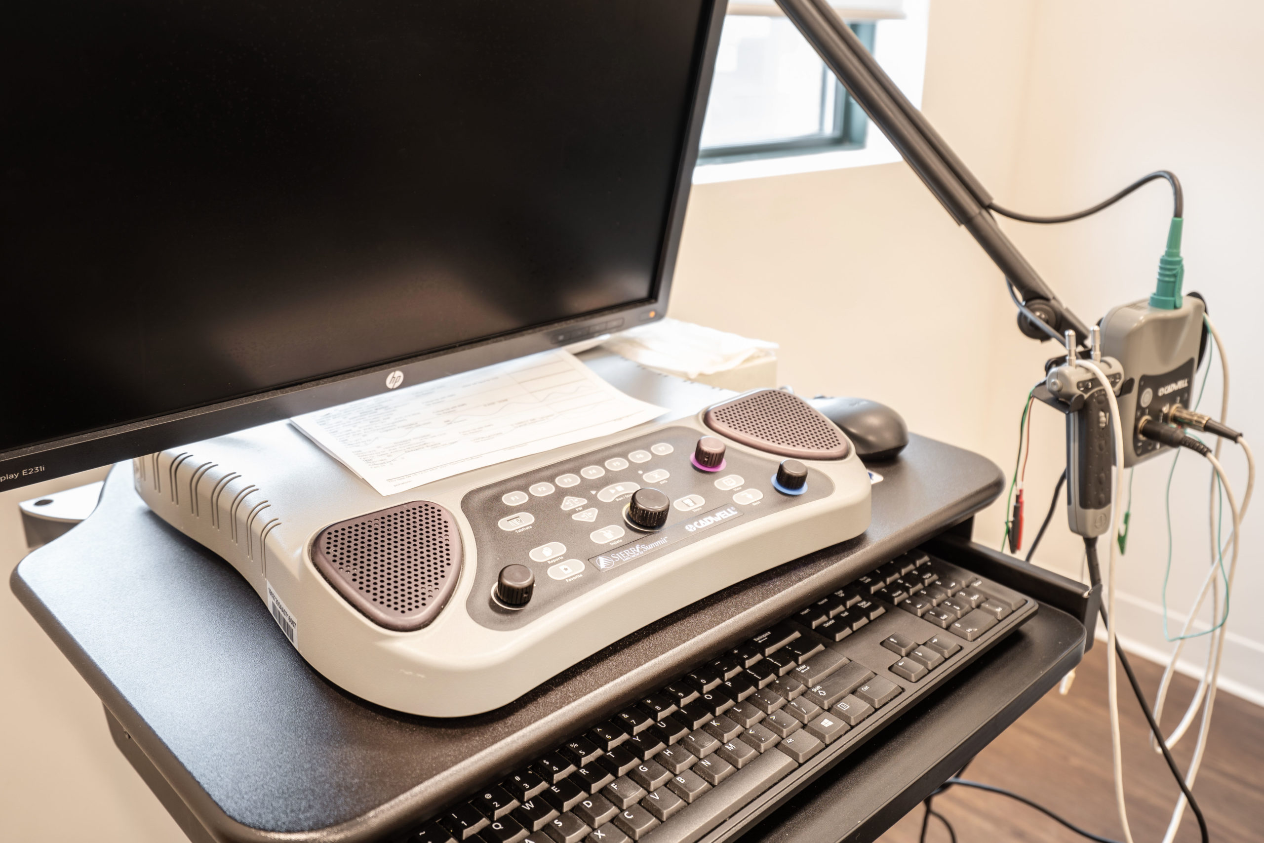
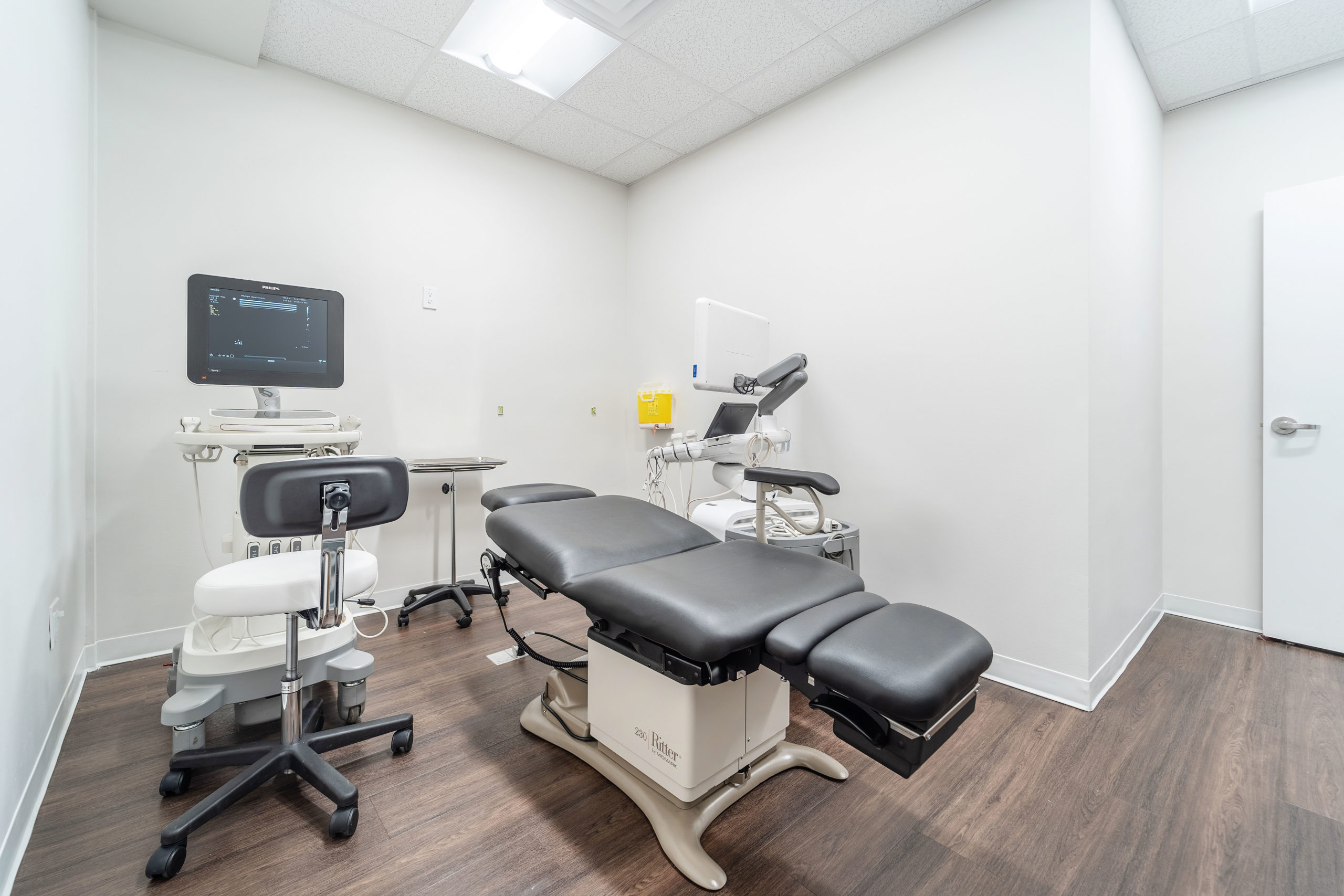
Nerve conduction studies (NCS) and electromyography (EMG) are often performed in conjunction to assess the functions of muscles and nerves. During NCS, small electrical impulses are sent into the muscles to assess the speed and strength of electric nerve signals travelling between two or more points, which are detected by surface electrode stickers on the skin. During needle EMG, a small needle is inserted into the muscle to detect the activity within the muscle and the related nerves. A specialized neurologist will interpret the signals and numerical values and share the results on the same day of the studies. Localized Neuromuscular ultrasound may also be performed to characterize the anatomy of a symptomatic site on the body and to assist with diagnoses.

EEG

An electroencephalogram (EEG) detects electrical activity in the brain. While it is one of the main diagnostic tests for epilepsy and seizure disorders, it can also provide information for other brain disorders. EEGs are safe and painless with no side effects. Small, metal discs (electrodes) will be placed on your scalp to capture brain wave activities. A specialized neurologist will then interpret the recording and formulate a report. To help in finding certain types of electrical activity in the brain, you may have to be asleep during the recording – this is called a sleep-deprived EEG. You may be asked not to sleep at all the night before the test or to sleep less by going to bed later and getting up earlier than usual. A routine procedure for an adult takes approximately 60 minutes. For children, the test will last approximately 90 minutes. Sleep-deprived testing also lasts approximately 90 minutes. We offer routine EEG with Video, sleep-deprived EEG, as well as 2-hour and 4-hour prolonged EEG.

Ultrasound

A noninvasive, diagnostic medical procedure that uses high frequency sound waves to produce dynamic visual images of organs, tissues, or blood supply inside the body. Carotid Doppler. Another noninvasive exam of the carotid and vertebral arteries of the neck, this test provides crucial information about blood flow to the brain. This diagnostic procedure is typically used to help determine risk of diseases such as stroke. Ultrasound is a procedure that examines the blood flow and detects blockages inside the arteries via sound waves, thereby determining the risk of stroke. We offer carotid doppler, 3D carotid plaque characterization, vascular ultrasound, and giant cell arteritis (GCA) ultrasound.

Vestibular Test Battery

Used to diagnose patients with balance or dizziness problems, vestibular tests are designed to evaluate the function and structure of the inner ear and/or brain. Electro-/Video-nystagmography (ENG/VNG) detects disorders in the peripheral vestibular system by placing electrodes around the eyes and using goggles with video cameras to monitor eye movements. Tests are usually administered in a low-light environment to evaluate signs of some vestibular and/or neurological dysfunctions. A caloric test, which changes the temperature within the ear canal as a stimulation to the vestibular system, can also be administered as part of the test battery. In addition to ENG/VNG with calorics, we also offer vHIT, VEMP, ECochG with ABR, and rotational tests.

Neuro-Ophthalmologic Tests

You will probably have your pupils dilated during the visit. The eye drops last about 4 hours and will make things look bright and blurry up close. Have someone else drive you to the appointment and bring your sunglasses. Copy: Visual field test measures all regions of your sight, including your peripheral vision. Fundus photography is an imaging study of the retina for evaluating Neuro-ophthalmologic disorders. This test is superior to the information your doctor can obtain with bedside fundoscopy. Optical coherence tomography (OCT) provides an even higher resolution scan of the retina for examining the anatomy of the retina and optic nerve (ganglion cell layer and retinal nerve fiber layer) in finer details. Subtle damage to the retinal layers is a early sign of brain or eye disease and can be missed in routine tests.

Cardiac Tests

Echocardiogram (Echo) is a cardiac ultrasound that detects dysfunctions within the heart via sound waves. Echo cardiograms with bubble study and contrast is a core test when evaluating for causes of stroke or TIA. Ask your doctor about contrast ECHO when routine echo is normal and no other cause of stroke has been identified. Holter monitoring is a portable electrocardiogram (ECG) that monitors electrical activity of the heart for a prolonged period of time. atrial fibrillation is a common cause of blood clots and stroke and requires prologed monitoring of the heart to detect as this sneaky rythym is seldom present continously. We offer echocardiograms with bubbles or contrast, Holter monitoring (48-hour, 72-hour, 14-day, and 28-day), as well as ambulatory blood pressure monitoring as part of either the stroke or syncope investigative pathway. In selected patients who have had an Embolic stroke of Uncertain Source a prolonged implantable device may be recommended for long term monitoring.

Home Sleep Study

Polysomnography, or commonly called sleep study, is a comprehensive set of recordings that monitors the brain waves, blood oxygen level, heart rate, breathing, and movement. This test primarily focuses on diagnosing obstructive sleep apnea (OSA). We offer level 3 home sleep studies: A home sleep study measures how well you sleep and the results from this test can determine if you have sleep apnea. These tests use a small home sleep recorder worn overnight while you sleep in the comfort of your own home. A fellowship certified sleep physician interprets the sleep test results to determine if you have sleep apnea.

Neuroradiology

Computerized tomography (CT) produces a series of cross-sectional X-ray images of the bones, blood vessels, and soft tissues. Magnetic resonance imaging (MRI) provides high-definition images of the organs and tissues in the body using a magnetic field and radio waves. While we don’t currently offer CT/MRI imaging at our facility, we are able to provide a second expert Neuro-radiologist opinion on interpreting raw CT/MRI imaging with special attention on the integrity of central and peripheral nervous systems. In addition, we have partnerships to be able to offer private pay and third party MRI and CT for non-OHIP requests.

Computerized neuropsychology

The cost of Neuropsychologic testing has rapidly increaded after this invaluable service was delisted from the OHIP schedule of benefits. At MINDS we are partnered with Cambridge Brain sciences - an innovative online Neuropsychology testing platform developed by leading neuropsychologists at Cambridge university. neuropsychology services for patients experiencing neuro-cognitive (related to memory, thinking and attention) and emotional difficulties that may result from a variety of conditions affecting the brain such as: Concussions Chronic headaches/migraine Seizure disorders or sleep disorders Psychiatric disorders such as depression and anxiety Alzheimer’s disease, Parkinson’s disease Strokes, brain tumors

FAQs

These are the most common questions we get about preparing for the EMG and EEG tests.

What happens during the EMG test?

The EMG test consists of two parts:

  • In the nerve conduction study, electrodes are placed on the skin and a safe level of electrical stimulation is sent through the nerve, causing the muscles in your arm or leg to contract. The sensation you will feel is similar to a static electric shock. Patients wearing pacemakers are not at risk, since this current is not strong enough or close enough to the pacemaker to interfere with its operation.
  • In the second part, the EMG, a tiny pin electrode is inserted under the surface of the skin into the muscle. Electrical stimulation will indicate if there is damage to a specific nerve, or if the problem involves the muscle itself.

This test is mildly uncomfortable but is specially designed to not be painful. It will take between 30 to 60 minutes.

How should I prepare for my NCS/EMG appointment?

  • Do not apply cream/lotion on your arms and legs prior to the appointment.
  • Wear loose-fitting clothes. You may also be asked to wear a gown during the testing.
  • Take your medications as usual with the exception of Mestinon, if applicable.

How should I prepare for my EEG appointment?

Preparation may vary according to the type of EEG testing or physician’s orders. In general:

  • Your hair should be clean and dry with no hair products (hairspray or gel).
  • Remove glue-on or sew-on wigs, braids, or hair extensions before your appointment.
  • Take medications as normal with the exception of sedatives, such as Ativan.
  • No caffeine intake (including coffee, tea, energy drink, and chocolate) before your appointment.
  • Wear comfortable and loose-fitting clothing.
  • You may bring a blanket to increase your comfort during the test.
  • You may bring a hat and/or a hairbrush/comb.
  • Allow for 1-1.5 hours in your schedule for an EEG appointment.

For sleep-deprived EEG’s:

  • This test is similar to a regular EEG, as described above, except that you will be asked to achieve 4 fewer hours of normal sleep or as directed by your provider prior to your exam time.
  • Do not eat or drink anything containing caffeine between midnight and the time of your test.
  • A sleep-deprived EEG can take up to 1.5-2 hours.
  • After the test, it is recommended that you be accompanied and do not drive.
  • Children being tested should be kept awake later than normal the night before and awakened a few hours earlier than usual. This unusual sleep schedule, along with keeping the child from napping, often helps the child lie still or fall asleep during testing.